Angiography Pack

| pcs | |
| 150x300 cm Femoral Angiography Drape | 1 |
| 100x150 cm Table Cover | 1 |
| 100x200 cm Drape Sheet | 1 |
| Fluoroscopy Cover | 1 |
| 40 x 40 cm Towel | 2 |
| Solution Bowl 250 cc. | 1 |
| 100x100 cm Wrapping Paper | 1 |

| pcs | |
| 150x300 cm Femoral Angiography Drape | 1 |
| 100x150 cm Table Cover | 1 |
| 100x200 cm Drape Sheet | 1 |
| Fluoroscopy Cover | 1 |
| 40 x 40 cm Towel | 2 |
| Solution Bowl 250 cc. | 1 |
| 100x100 cm Wrapping Paper | 1 |

Bionano Data Solutions™ includes a complete suite of hardware and software for end-to-end experiment management, analysis and bioinformatics processing, along with convenient web-based management and monitoring tools.
Automate data analysis: monitor and manage Saphyr remotely
Bionano Access™, your web-based hub for Saphyr operations, provides all the software you need for experiment management and Bionano optical genome mapping in one place.
Reduce infrastructure costs and increase your optical genome mapping capacity
Bionano Compute On Demand is a pay-per-use solution accessible through Bionano Access web server for your Bionano Solve operations. Compute On Demand simplifies the way you perform genome assembly, hybrid scaffolding, and structural variant analysis, without the need for any additional infrastructure, giving you the flexibility and scalability your experiment deserves. Advantages include:
Bionano Compute on Demand requires Bionano Access 1.6.1 which is available on our Software Downloads page.
For more information, download the Bionano Compute On Demand brochure.
| Human de Novo Assembly | 1 | Calculated | Max tokens computed based on input molecules. 2 Tbp limit (down sampled to 250x at runtime) |
| Non-Human or No Reference de Novo Assembly | 1 | 50 | Below 300 Gbp |
| Non-Human or No Reference de Novo Assembly | 10 | 150 | 300 - 500 Gbp |
| Non-Human or No Reference de Novo Assembly | 20 | 250 | 500 - 1000 Gbp |
| Non-Human or No Reference de Novo Assembly | 30 | 400 | 1000 - 2000 Gbp |
| Non-Human or No Reference de Novo Assembly | 40 | 500 | 2000 - 3000 Gbp (Limit Increased) |
| Non-Human or No Reference de Novo Assembly | 50 | 650 | 3000 - 4000 Gbp (New Tier) |
| Scaffold | 1 | 30 | |
| Two Enzyme Hybrid Scaffold | 1 | 30 | |
| Variant Annotation (Single) | 1 | 3 | |
| Variant Annotation (dual or trio) | 1 | 10 | |
| Bnx Merge | 1 | 2 | |
| Filter Bnx | 1 | 2 | |
| MQR | 1 | 2 | |
| SV Merge | 1 | 5 | |
| Alignment | 1 | 4 | |
| Rare Variant Analysis | 3 | 25 | 0 - 2000 Gbp |
| Rare Variant Analysis | 5 | 50 | 2000 - 6000 Gbp (New Tier) |
| Bionano Enfocus FSHD Analysis | 1 | 5 | New Operation |
| OPERATION | MIN TOKENS | MAX TOKENS | NOTES |
|---|
*Number of tokens required dependent on the data quality and coverage
Enhanced data processing power for high-speed optical genome mapping
The Saphyr Compute Server offers cluster-like performance in an affordable, compact solution. Capable of performing multiple simultaneous analyses and sustaining continuous throughput, the Saphyr Compute Server allows you to spend less time waiting for data so you can focus on investigating results.
Additional power for Saphyr
The Bionano Compute Server is a stand-alone, secondary compute server that offers the ability to perform automated genome assembly, hybrid scaffolding and structural variant calling in parallel with the Saphyr Compute Server. Additional Bionano Compute Servers can be added for increased processing power.
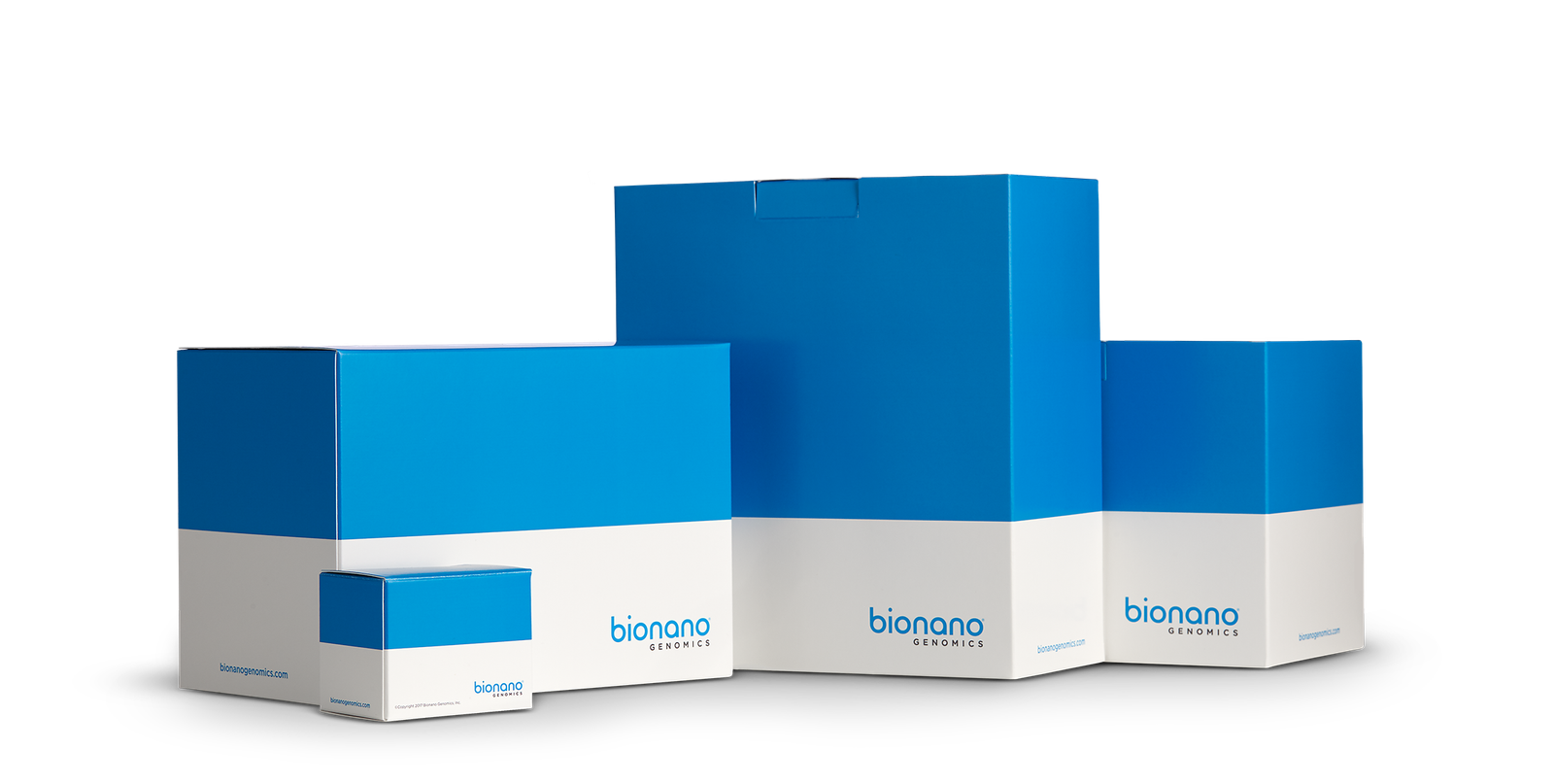
Bionano Prep Kits™ provide the critical reagents and protocols needed to extract and label high molecular weight (HMW) DNA for use on the Irys® and Saphyr™ systems. Bionano kits are optimized for performing Bionano optical genome mapping applications on a variety of sample types.
Bionano optical genome mapping begins with the isolation of HMW DNA
Bionano sample kits are optimized for isolating and purifying HMW DNA in a process that is gentler than existing HMW DNA extraction methods. The resulting purified DNA, several megabases in length, is optimal for use with Bionano systems. Each Bionano Prep Kit allows you to perform 5-10 HMW DNA preps. Bionano kits and protocols enable the extraction of HMW DNA from a variety of sample types including soft or fibrous:
| NEW! 90057 |
Bone Marrow Aspirate(BMA) DNA Isolation kit – 10 reactions | For use with heparin-collected bone marrow aspirates (BMA). Compatible with Bionano DLS DNA Labeling Kit only. First-time users must order Bionano Prep SP Magnetic Retriever (#80031). |
| NEW! 80038 |
SP Tissue and Tumor DNA Isolation kit – 10 reactions | For use with human or animal tissue or tumor. Compatible with Bionano DLS DNA Labeling Kit only. First-time users must order Bionano Prep SP Magnetic Retriever (#80031). |
| 80030 | SP Blood & Cell Culture DNA Isolation Kit – 10 reactions | For use with EDTA-collected blood and mammalian cell cultures. Compatible with Bionano DLS DNA Labeling Kit only. First-time users must order Bionano Prep SP Magnetic Retriever (#80031). |
| 80002 | Animal Tissue DNA Isolation Kit - 10 reactions | For use with soft or fibrous animal tissue. Order with Bionano DLS DNA Labeling Kit or with Bionano NLRS DNA Labeling Kit. |
| 80004 | Blood and Cell Culture DNA Isolation Kit - 10 reactions | For use with blood and cell cultures. Order with Bionano DLS DNA Labeling Kit or with Bionano NLRS DNA Labeling Kit. |
| 80003 | Plant DNA Isolation Kit - 5 reactions | For use with most plant tissues. Order with Bionano DLS DNA Labeling Kit or with Bionano NLRS DNA Labeling Kit. |
| 80005 | DLS DNA Labeling Kit - 10 reactions | Direct Label and Stain Kit for use with Bionano Animal Tissue, Plant, or Blood and Cell Culture DNA Isolation Kits. Not required for using the Bionano Prep Kit. |
| 80001 | NLRS DNA Labeling Kit - 10 reactions | Nicking, Labeling, Repairing and Staining Kit for use with Bionano Animal Tissue, Plant, or Blood and Cell Culture DNA Isolation Kits. Not required for using the Bionano Prep Kit. |
| CATALOG # | COMPONENT | PRODUCT DESCRIPTION |
|---|
Bionano labeling reagents are optimized for applications on Bionano optical genome mapping systems
Starting with HMW DNA purified using the appropriate Bionano prep kit, fluorescent labels are attached to specific sequence motifs. The result is uniquely identifiable genome-specific label patterns that enable de novo map assembly, anchoring sequencing contigs and discovery of structural variations starting at 500 bp.

Built using proprietary Nanochannel technology, Bionano Chips for the Saphyr® and Irys® systems linearize DNA, enabling high-speed, high-throughput optical genome mapping and structural variation detection for a variety of applications including human and clinical research.
The Bionano Saphyr Chip® and Irys Chip® utilize hundreds of thousands of massively parallel NanoChannels per flow cell that linearize long DNA molecules allowing Bionano systems to directly image your samples.
Inside the chip’s flow cell, a gradient of micro- and nano-structures, upstream of the chip’s NanoChannels, gently unwinds and guides DNA into the NanoChannels. Saphyr Chip’s NanoChannels allow only a single linearized DNA molecule to travel through while preventing the molecule from tangling or folding back on itself.
By leveraging mature semiconductor manufacturing technology, Bionano delivers robust performance and quality. The breakthrough Saphyr Chip combines with the Bionano Direct Label and Staining (DLS) technology to provide you with the highest sensitivity possible for structural variant detection starting at 500 bp, along with genome maps up to chromosome-arm-length.
| Flowcells | 3 | 2 | 2 |
| Total Output (human sample/flowcell) |
5 Tbp* | 1300 Gbp* | 10 Gbp / 50 Gbp |
| Hardware Compatibility | Saphyr Instrument (Part #60325) |
Saphyr Instrument (Part #60239) |
Irys Instrument |
| Catalog Number | 20366 | 20319 | 20249 |
| Specification Sheet | Download Here | Download Here | N/A |
| FEATURES | SAPHYR CHIP G2.3 | SAPHYR CHIP G1.2 | IRYS CHIP |
|---|
Specifications based on Bionano-supplied NA12878 control sample prepared with the Bionano Prep SP kit.
For non-control samples, performance may vary due to a number of factors, including quality of starting material.
*Bionano supplied control sample only

Optical genome mapping using Saphyr® reveals what’s missing in your research. Rapidly identify genome variation like never before with the high-throughput Saphyr system.
Resolve large-scale structural variations missed by next-generation sequencing (NGS) systems
Large structural variations are responsible for many diseases and conditions, including cancers and developmental disorders. Optical genome mapping with Saphyr detects structural variations ranging from 500 bp to megabase pairs in length and offers assembly and discovery algorithms that far outperform sequencing-based technologies in sensitivity.
For mosaic samples or heterogeneous cancer samples, Saphyr detects all types of structural variants down to 1% variant allele fraction. Saphyr provides this performance typically with a false positive rate of less than 2%. Saphyr also calls repeats and complex rearrangements.
Detect large-scale structural variations for genetic disease, cancer, and cytogenomics.
Rapid optical genome mapping using whole-genome imaging for human research applications
Saphyr features enhanced optics with adaptive loading of DNA utilizing machine learning. The Saphyr Instrument and high-capacity Saphyr Chip® combine to deliver genome maps at the speed and scale your research demands.
Optical genome mapping automation features and intelligent sample preparation simplify the process
Saphyr optical genome mapping offers automated features that minimize hands-on time.
Optical genome mapping automation features and intelligent sample preparation simplify the process
Saphyr runs each genome in its own flow cell. Each Saphyr chip holds three flow cells, and 100x coverage of 3 human genomes is collected in less than 6 hours. That means 12 human samples at 100x coverage per day, or up to 96 per week.
For cases requiring higher coverage, like mosaic samples or heterogeneous tumors, the run can be extended up to 24 hours to collect as much as 400x coverage of a human genome for each of the three flowcells.
Remote quality control ensures maximum performance at all time
The Saphyr Assure Service provides an optional (opt-in required) automated health monitoring feature that continuously inspects data quality and instrument performance. Performance issues are detected before data quality and performance are impacted allowing Bionano Support to proactively repair the system with minimal downtime.
Automate data analysis—monitor and manage the Saphyr Instrument remotely
The Bionano suite of hardware and software for Saphyr streamlines your workflow and reduces time to result.
Extract high-molecular-weight DNA for human, plant and animal genome mapping
The Bionano Prep DLS DNA labeling kit provides all the enzymes, dyes and buffers you need to label non-amplified DNA for Saphyr. Controls for validating the labeling biochemistry are also provided.
*Compatible only with Saphyr System #90023 (Saphyr Instrument #60325)
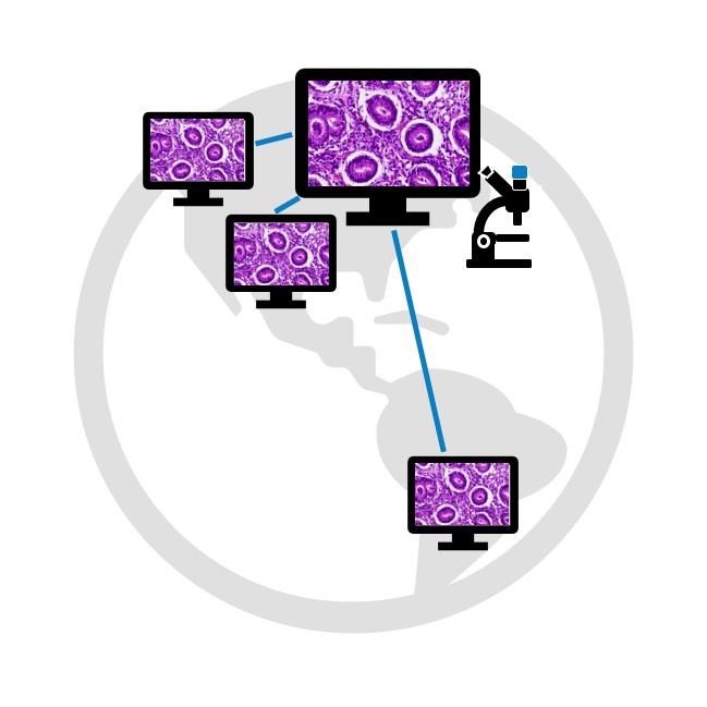
Bridge distances and keep working from anywhere.
Save time without taking any risk.

6 months starter package
purchase additional months depending on your needs
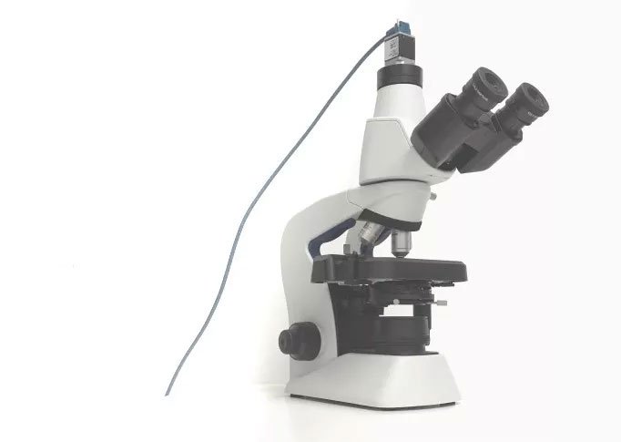
For upgrade of microscopes with a trinocular head.
Microvisioneer manualWSI Scanning Software (does not include fluorescence scanning)
Microvisioneer InstaSlide Scan Sharing Software
Camera suitable for manual scanning
Camera adapter
Guaranteed shipping within 2-3 business days
Share digital slides within seconds with anyone, anywhere.
View slides from any distant location.
Your telemicroscopy solution.
Our software solutions are affordable and immediately available.
Used in 50+ countries worldwide.
Everyone receiving the URL can view the virtual slides now. Nothing more than a web browser is needed.
The scans can be viewed as a zoomable website on any device such as computers, tablets or smartphones.
The capacity of the InstaSlide virtual slides server is unlimited. You can share hundreds of slides within seconds.
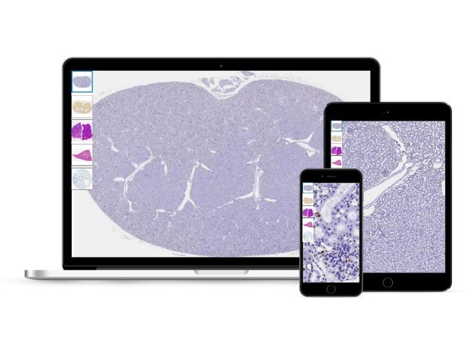
Generally, all digital images in svs or tiff file format can be shared.
InstaSlide of course works perfectly with Microvisioneer's manual scanning software manualWSI.
As manual slide scanning is much more affordable than motorized scanning, a telemicroscopy workflow can be easily set up anywhere very quickly and without financial risks.
Upgrade your microscope to a manual slide scanner.
Start creating virtual microscope slides now for any digital pathology application.
Our software solutions are affordable and immediately available.
Used in 50+ countries worldwide.
Now also available as
Fluorescence-Edition for fluorescence slide scanning!
Convert your existing microscope with a suitable camera and the Microvisioneer manualWSI scanning software to a manual microscope slide scanner.
Camera mounting and software installation is extremely easy and can be done within 5 min.
A simple microscope upgrade - your ticket to virtual microscopy.
Digitize your glass slides:
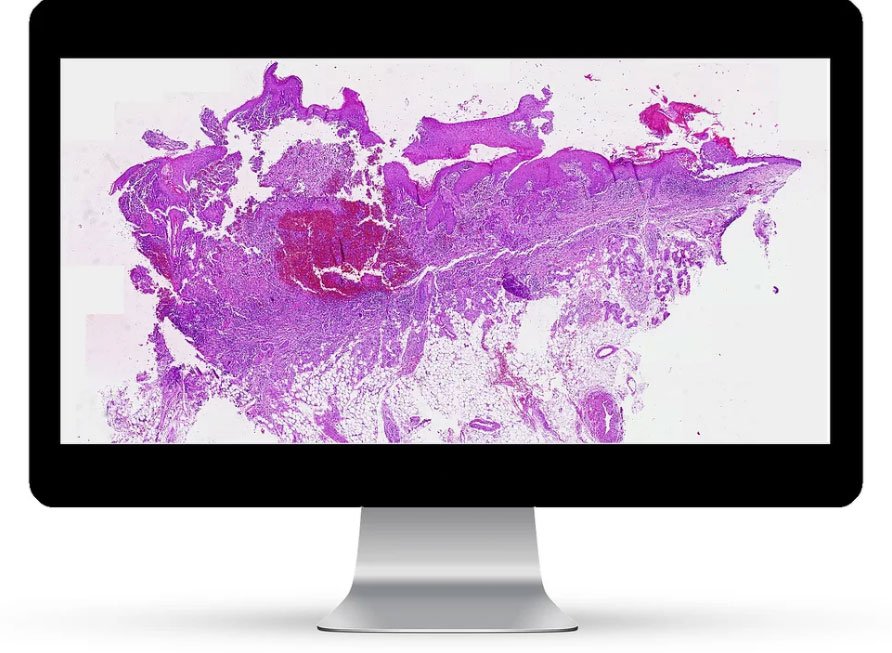
Select any desired objective and scan high-quality zoomable whole slide images or representative subregions of the specimens, using your microscope as whole slide image scanner.
Scanning is intuitive and smooth. Real time image stitching is seamless.

Microvisioneer virtual slides can be viewed and analyzed by the major available viewers and image analysis tools. Many of them are freely available, such as Sedeen or QuPath. The manual scanner can be integrated into any digital pathology workflow.
For sharing the images, simply use cloud-based systems or find out more about the Microvisioneer InstaSlide virtual slide sharing solution
IT GIVES A CLEAR OVERVIEW. IT IS VERY FAST. IT HAS A GREAT RESOLUTION. IT IS EASY TO FOCUS WHEREEVER YOU NEED IT.
A WORKING SCANNER THAT PRODUCES DIAGNOSTIC QUALITY IMAGES
I CAN USE MY EXISTING EQUIPMENT, NO NEED FOR A MAJOR CAPITAL INVESTMENT. MANY OF THE FEATURES ARE UNIQUE AND USEFUL.
UNPRECEDENTED SMOOTHNESS IN THE IMAGE STITCHING, WHICH IS IMPRESSIVE
PRACTICAL AND EASY USABILITY FOR OIL-BASED SCANNING
Microvisioneer's manual scanning approach combines digital progress and innovation with the versatility and flexibility of traditional microscopes. With the resulting manual microscope scanner, almost any type of slide can be digitized, no matter how exceptional.
For example, particularly thick slides such as brain tissue sections or uneven slides, which are often a problem for automated scanners, can be perfectly scanned.
Applications range from virtual slides pathology, digital histology, cytology, hematology, veterinary and plant pathology to geology and material science.
Typical brightfield, oil immersion, phase contrast and polarized light imaging are possible as well as fluorescence imaging with the newly available manualWSI-Fluorescence edition.
Explore a few selected applications in more detail in our use case section on this website.
You decide what you scan and at which objective lens magnification.
For research use only.