manualWSI software
Upgrade your microscope to a manual slide scanner.
Start creating virtual microscope slides now for any digital pathology application.
Our software solutions are affordable and immediately available.
Used in 50+ countries worldwide.
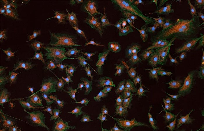
Now also available as
Fluorescence-Edition for fluorescence slide scanning!
Manual WSI
Watch a Quick Demo
The Manual Scanning Process
UPGRADE YOUR MICROSCOPE TO A MANUAL SCANNER
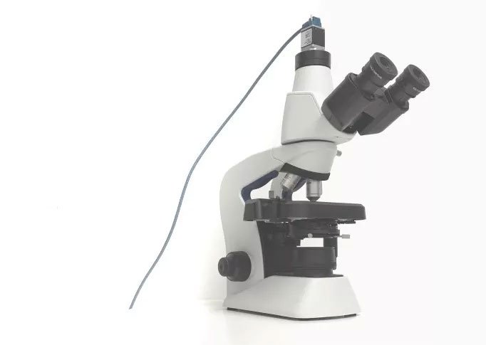
Convert your existing microscope with a suitable camera and the Microvisioneer manualWSI scanning software to a manual microscope slide scanner.
Camera mounting and software installation is extremely easy and can be done within 5 min.
A simple microscope upgrade – your ticket to virtual microscopy.
CREATE ZOOMABLE WHOLE SLIDE IMAGES…
Digitize your glass slides:
Select any desired objective and scan high-quality zoomable whole slide images or representative subregions of the specimens, using your microscope as whole slide image scanner.
Scanning is intuitive and smooth. Real time image stitching is seamless.

VIEW, ANALYZE, ARCHIVE OR SHARE THE VIRTUAL SLIDES
Microvisioneer virtual slides can be viewed and analyzed by the major available viewers and image analysis tools. Many of them are freely available, such as Sedeen or QuPath. The manual scanner can be integrated into any digital pathology workflow.
For sharing the images, simply use cloud-based systems or find out more about the Microvisioneer InstaSlide virtual slide sharing solution
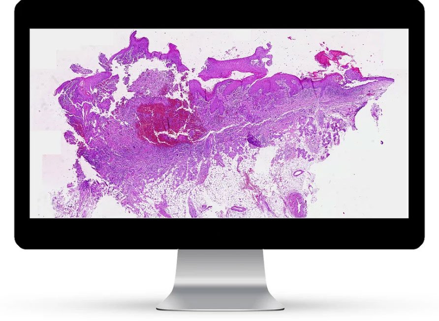
Zoom into Resulting Whole Slide Images!
Possibilities of Whole Slide Imaging
Benefits of Microvisioneer manualWSI
What Our Customers Say
IT GIVES A CLEAR OVERVIEW. IT IS VERY FAST. IT HAS A GREAT RESOLUTION. IT IS EASY TO FOCUS WHEREEVER YOU NEED IT.
A WORKING SCANNER THAT PRODUCES DIAGNOSTIC QUALITY IMAGES
I CAN USE MY EXISTING EQUIPMENT, NO NEED FOR A MAJOR CAPITAL INVESTMENT. MANY OF THE FEATURES ARE UNIQUE AND USEFUL.
UNPRECEDENTED SMOOTHNESS IN THE IMAGE STITCHING, WHICH IS IMPRESSIVE
PRACTICAL AND EASY USABILITY FOR OIL-BASED SCANNING
Applications
Microvisioneer’s manual scanning approach combines digital progress and innovation with the versatility and flexibility of traditional microscopes. With the resulting manual microscope scanner, almost any type of slide can be digitized, no matter how exceptional.
For example, particularly thick slides such as brain tissue sections or uneven slides, which are often a problem for automated scanners, can be perfectly scanned.
Applications range from virtual slides pathology, digital histology, cytology, hematology, veterinary and plant pathology to geology and material science.
Typical brightfield, oil immersion, phase contrast and polarized light imaging are possible as well as fluorescence imaging with the newly available manualWSI-Fluorescence edition.
Explore a few selected applications in more detail in our use case section on this website.
You decide what you scan and at which objective lens magnification.
For research use only.


